Poultry Scientists Develop 3D Anatomy Technique to Learn More About Chicken Vision

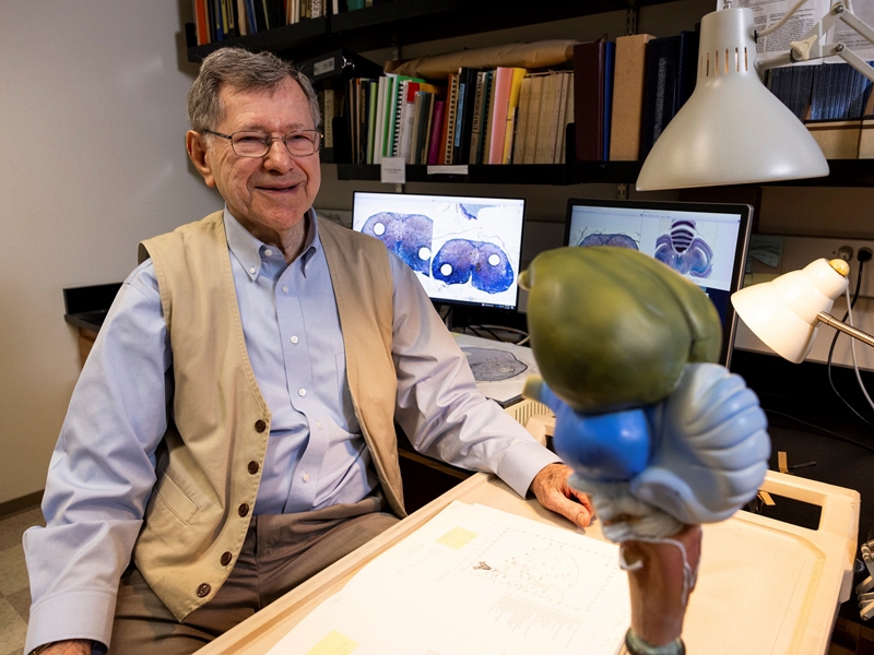
Wayne Kuenzel, professor of physiology and neuroendocrinology in the department of poultry science, explains diceCT imaging at his office in the Center of Excellence for Poultry Science at the University of Arkansas.
By John Lovett
University of Arkansas System Division of Agriculture
FAYETTEVILLE, Ark. – Poultry scientists with the Arkansas Agricultural Experiment Station are unraveling the complexities of bird brains and finding less expensive ways to do it.
The scientists mapped the intricate neurological pathways that control vision in chickens with detailed 3D models of the connections between the eyes and four regions of the brain. The research paper, titled "Mapping the avian visual tectofugal pathway using 3D reconstruction," was accepted for publication in the Journal of Comparative Neurology. A separate research paper was written on the thalamofugal pathway.
Wayne Kuenzel, professor of physiology and neuroendocrinology in the department of poultry science for the experiment station, said the technique is a less expensive way to create quality 3D images resembling magnetic resonance imaging, or MRI, technology. He also said the method will benefit teaching complex anatomy and expand the tools of animal science researchers. The experiment station is the research arm of the University of Arkansas System Division of Agriculture.
"What is important about this technique is that it is a straightforward procedure, and it is not expensive," Kuenzel said. "I am sure it will gain importance over time and attract a greater audience."
Parker Straight, principal author of the research publication, pursued a Master of Science degree under Kuenzel within the Division of Agriculture's Center of Excellence for Poultry Science. Straight has gone on to work as a clinical research associate and avian neuroanatomy research consultant with Kuenzel as they update "Stereotaxic Atlas of the Brain of the Chicken," a book detailing the anatomy of chicken brains first published by Kuenzel in 1988.
Paul Gignac, associate professor of cellular and molecular medicine with the University of Arizona College of Medicine–Tucson, was a member of Parker's thesis committee and a co-author of the 3D imaging study.
"It's not only a quality research paper but will also be helpful in teaching," Kuenzel said of Straight's work. "The tectofugal visual pathway has four critical neural structures in four different brain regions. Diagramming them in 3D enables one to see the entire pathway in one image and therefore should enable the learning of the entire pathway more rapidly and perhaps more permanently."
To create the new 3D imaging, Straight said they combined a conventional imaging method called histochemistry with a newer imaging method known as diceCT, which stands for "diffusible iodine-based contrast-enhance computer tomography."
Histochemistry uses chemical reagents like dyes to stain tissue and allow it to undergo image analysis. DiceCT is like an MRI, Straight explained, but instead of using a large magnet and radio waves, it uses iodine to stain the tissue so that a viewer can see groups of cells among fiber tracts. DiceCT uses x-ray scans to "digitally" slice the biological subject being studied.
Straight, Gignac and Kuenzel modeled the tectofugal pathway, the primary visual pathway in chickens, by combining the technologies with data reconstruction computer programs such as Brainmaker, Avizo and Blender. Kuenzel said Gignac has been instrumental with many scientists in developing and describing the diceCT procedure.
The iodine used in diceCT is not permanent and can be removed from the sample tissue without damaging or distorting the tissue, which is important for the integrity of the 3D imaging, Straight added.
"With the method being cheaper, it allows it to be accessible to many more researchers who often may not consider pursuing the use of MRI due to its cost or availability," Straight said.
Kuenzel noted that the research paper could also "broaden the diversity of scientists who might add the Journal of Comparative Neurology to their list of journals to review more regularly."
Why it's important
Straight said the hybrid method of 3D scanning can be used to study neurobiology at a large scale, such as brain region morphology, and at a more detailed scale, such as looking at a single neurological pathway. One example of the technology's potential use would include assessing changes or lesion patterns at various stages of a disease.
Other examples, he said, may include long-distance neuron tracing without cutting the connection, as well as comparing structural differences and how they relate to different behavioral patterns.
"The list is quite long in terms of how this method can be proven beneficial to research," Straight said. "I hope this study will prompt more investigations of animal neurobiology using 3D methods and how it compares to neurobiology of humans."
Straight noted that if a researcher wanted to implement the exact imaging pipeline they used, the bird would have to be euthanized. However, the diceCT portion of the imaging method can be done in live animals if they are sufficiently sedated so that a researcher can capture a clean 3D scan.
The research was supported in part by grants from the University of Arkansas' Chancellor's Innovation Fund and the Arkansas Biosciences Institute.
To learn more about Division of Agriculture research, visit the Arkansas Agricultural Experiment Station website: https://aaes.uada.edu. Follow on Twitter at @ArkAgResearch. To learn more about the Division of Agriculture, visit https://uada.edu/. Follow us on Twitter at @AgInArk. To learn about extension programs in Arkansas, contact your local Cooperative Extension Service agent or visit www.uaex.uada.edu.
About the Division of Agriculture: The University of Arkansas System Division of Agriculture's mission is to strengthen agriculture, communities, and families by connecting trusted research to the adoption of best practices. Through the Agricultural Experiment Station and the Cooperative Extension Service, the Division of Agriculture conducts research and extension work within the nation's historic land grant education system. The Division of Agriculture is one of 20 entities within the University of Arkansas System. It has offices in all 75 counties in Arkansas and faculty on five system campuses. The University of Arkansas System Division of Agriculture offers all its Extension and Research programs and services without regard to race, color, sex, gender identity, sexual orientation, national origin, religion, age, disability, marital or veteran status, genetic information, or any other legally protected status, and is an Affirmative Action/Equal Opportunity Employer.
Topics
Contacts
John Lovett, project/program specialist
Agricultural Communication Services
479-763-5929,
jl119@uark.edu
Headlines
PetSmart CEO J.K. Symancyk to Speak at Walton College Commencement
J.K. Symancyk is an alumnus of the Sam M. Walton College of Business and serves on the Dean’s Executive Advisory Board.
Faulkner Center, Arkansas PBS Partner to Screen Documentary 'Gospel'
The Faulkner Performing Arts Center will host a screening of Gospel, a documentary exploring the origin of Black spirituality through sermon and song, in partnership with Arkansas PBS at 7:30 p.m. Thursday, May 2.
UAPD Officers Mills and Edwards Honored With New Roles
Veterans of the U of A Police Department, Matt Mills has been promoted to assistant chief, and Crandall Edwards has been promoted to administrative captain.
Community Design Center's Greenway Urbanism Project Wins LIV Hospitality Design Award
"Greenway Urbanism" is one of six urban strategies proposed under the Framework Plan for Cherokee Village, a project that received funding through an Our Town grant from the National Endowment for the Arts.
Spring Bike Drive Refurbishes Old Bikes for New Students
All donated bikes will be given to Pedal It Forward, a local nonprofit that will refurbish your bike and return it to the U of A campus to be gifted to a student in need. Hundreds of students have already benefited.


